Difference between revisions of "Ultrasound modulated optical tomography using contrast agents"
Qimei Zhang (talk | contribs) (Created page with " ==Ultrasound mediated luminescence tomography using contrast agents== ==Introduction:== Macroscopic fluorescence imaging and bioluminescence imaging are important molec...") |
Qimei Zhang (talk | contribs) (→Projects:) |
||
| Line 9: | Line 9: | ||
1) The mechanisms of USMLT are investigated. This is achieved through incorporate the effects of ultrasound modulation to an open source tool NIRFAST for simulation of diffusely propagating light. The modulated fluorescent signal is successfully calculated through the finite element method. The effects of four factors (reduced optical scattering coefficient, optical absorption coefficient, refractive index, and luciferase concentration) on the depth of light modulation are compared. | 1) The mechanisms of USMLT are investigated. This is achieved through incorporate the effects of ultrasound modulation to an open source tool NIRFAST for simulation of diffusely propagating light. The modulated fluorescent signal is successfully calculated through the finite element method. The effects of four factors (reduced optical scattering coefficient, optical absorption coefficient, refractive index, and luciferase concentration) on the depth of light modulation are compared. | ||
| − | + | [[File:Qimei_Zhang_USMBLTSimulation.pdf]] | |
| − | |||
2) The fluorophore-labelled microbubbles are investigated as a contrast agent. Fluorophore-labeled MBs can be tuned to be self-quenched at optimized concentration of fluorophore per MBs. With application of US, fluorophore-labeled microbubbles oscillate and thus result in the intermolecular spacing alteration. Such intermolecular oscillation could lead to a higher modulation of fluorescence signal of fluorophore-labeled MBs due to the fluorescence recovery of the self-quenched fluorophore. | 2) The fluorophore-labelled microbubbles are investigated as a contrast agent. Fluorophore-labeled MBs can be tuned to be self-quenched at optimized concentration of fluorophore per MBs. With application of US, fluorophore-labeled microbubbles oscillate and thus result in the intermolecular spacing alteration. Such intermolecular oscillation could lead to a higher modulation of fluorescence signal of fluorophore-labeled MBs due to the fluorescence recovery of the self-quenched fluorophore. | ||
| − | + | [[File:Qimei_Zhang_schameticFMB]] | |
| + | |||
| + | [[File:Qimei_Zhang_10pulse0dot75um100kPa.pdf]] | ||
3) Pyrene labelled liposomes are investigated as another contrast agent. Pyrene has a well-known concentration dependant excimer formation characteristics. In this project pyrene are labelled in the lipid bilayer of nanoscale liposomes and the excimer fluorescence intensity upon application of ultrasound is monitored. | 3) Pyrene labelled liposomes are investigated as another contrast agent. Pyrene has a well-known concentration dependant excimer formation characteristics. In this project pyrene are labelled in the lipid bilayer of nanoscale liposomes and the excimer fluorescence intensity upon application of ultrasound is monitored. | ||
| − | |||
| − | |||
| − | |||
| − | |||
4) Donor-acceptor labelled liposomes are also investigated as a contrast agent based on fluorescence resonance energy transfer (FRET). This contrast agent works at near-infrared wavelength range which is better for deep tissue imaging. | 4) Donor-acceptor labelled liposomes are also investigated as a contrast agent based on fluorescence resonance energy transfer (FRET). This contrast agent works at near-infrared wavelength range which is better for deep tissue imaging. | ||
Publications: | Publications: | ||
Revision as of 23:20, 26 April 2016
Ultrasound mediated luminescence tomography using contrast agents
Introduction:
Macroscopic fluorescence imaging and bioluminescence imaging are important molecular imaging methods for deep tissue interrogations. However, the imaging depth and resolution are limited by strong tissue scattering and absorption. The hybrid technique Ultrasound modulated luminescence tomography (USMLT) has been investigated in recent years aiming to provide images with ultrasound resolution (< 1 mm) in deep tissue (>10 mm). In this technique, a focused ultrasound is used to modulate the source particle region (e.g. fluorophores) or the optical properties of a scattering medium and the modulated signal is detected. By scanning the US beam, images can be obtained with a resolution decided by the ultrasound focal zone. However, this technique has a limitation that the modulation depth (ratio of the modulated light with unmodulated light) is extremely low leading to a low SNR for the system. The aim of this project is thus to increase the modulation depth (or the US On-to-Off ratio) of USMLT.
Projects:
1) The mechanisms of USMLT are investigated. This is achieved through incorporate the effects of ultrasound modulation to an open source tool NIRFAST for simulation of diffusely propagating light. The modulated fluorescent signal is successfully calculated through the finite element method. The effects of four factors (reduced optical scattering coefficient, optical absorption coefficient, refractive index, and luciferase concentration) on the depth of light modulation are compared.

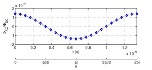
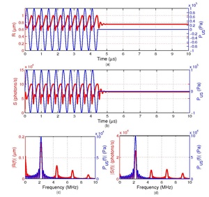
2) The fluorophore-labelled microbubbles are investigated as a contrast agent. Fluorophore-labeled MBs can be tuned to be self-quenched at optimized concentration of fluorophore per MBs. With application of US, fluorophore-labeled microbubbles oscillate and thus result in the intermolecular spacing alteration. Such intermolecular oscillation could lead to a higher modulation of fluorescence signal of fluorophore-labeled MBs due to the fluorescence recovery of the self-quenched fluorophore.
3) Pyrene labelled liposomes are investigated as another contrast agent. Pyrene has a well-known concentration dependant excimer formation characteristics. In this project pyrene are labelled in the lipid bilayer of nanoscale liposomes and the excimer fluorescence intensity upon application of ultrasound is monitored.
4) Donor-acceptor labelled liposomes are also investigated as a contrast agent based on fluorescence resonance energy transfer (FRET). This contrast agent works at near-infrared wavelength range which is better for deep tissue imaging.
Publications:
